Dermatome Map Lower Leg – When viewed on a body map, they appear like stacked discs. The dermatome pattern in the limbs is slightly different. This is due to the shape of the limbs as compared with the rest of the body. . This is a rare case of Varicella zoster virus (VZV) lumbosacral plexopathy in an 84-year-old women presenting with lower leg, and she can no longer drive a vehicle. The patient continues to .
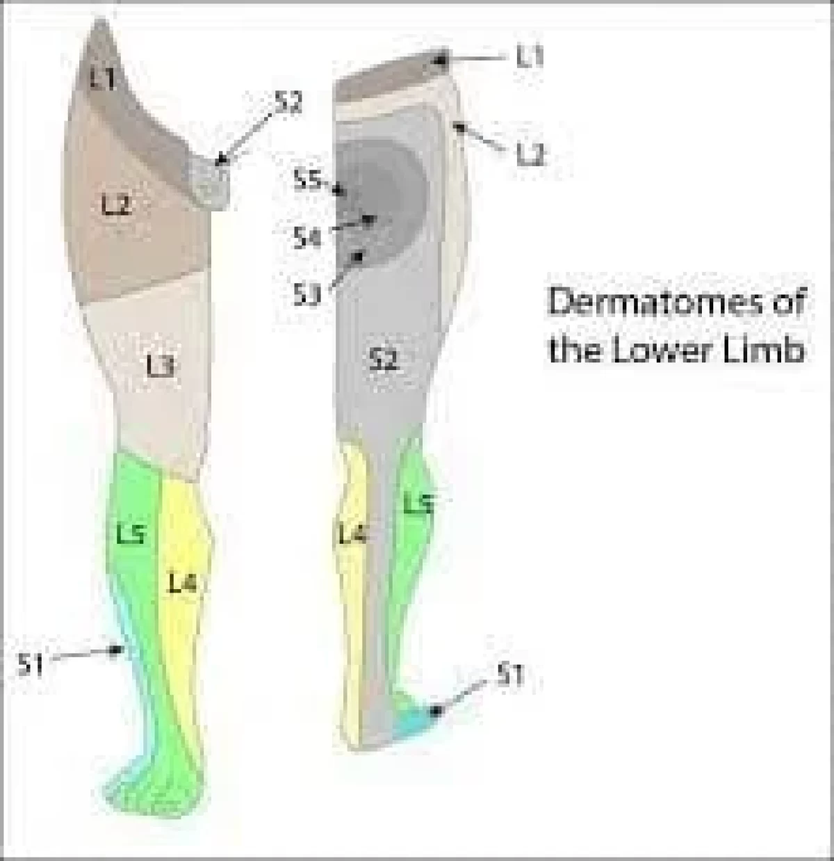
Dermatome Map Lower Leg
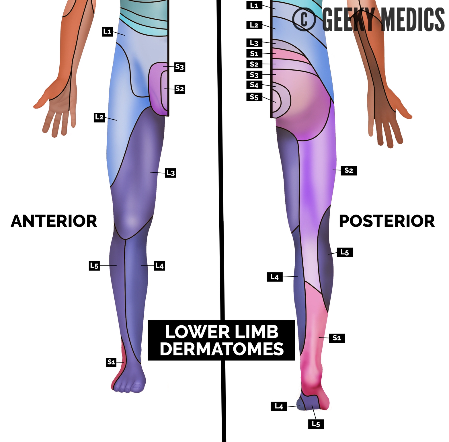
Source : geekymedics.com
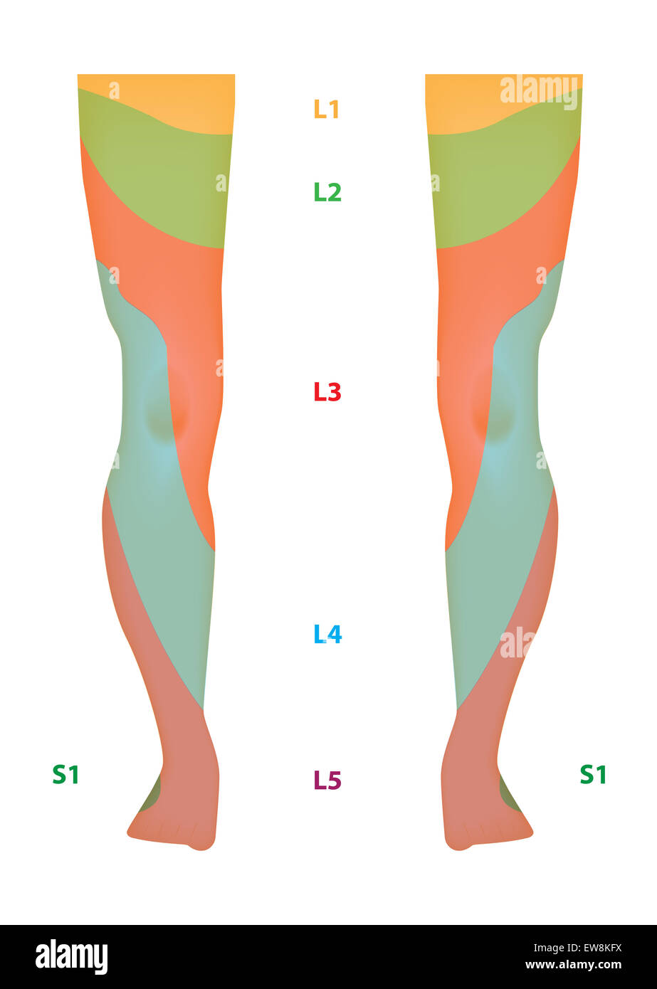
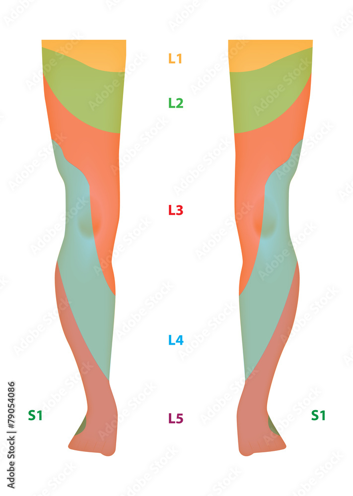
Dermatome Map of the Lower Limb Stock Vector | Adobe Stock

Source : stock.adobe.com
Dermatome Map of the Lower Limb Stock Photo Alamy
Source : www.alamy.com
The Lower Limb | SpringerLink
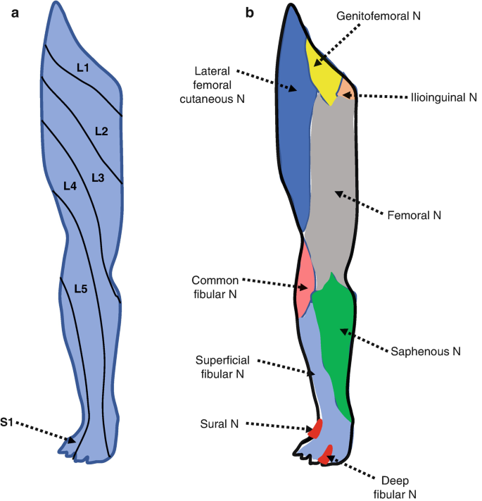
Source : link.springer.com
Dermatomes: Definition, chart, and diagram

Source : www.medicalnewstoday.com
Dermatomes Neurology Medbullets Step 1

Source : step1.medbullets.com
The Lower Limb | SpringerLink
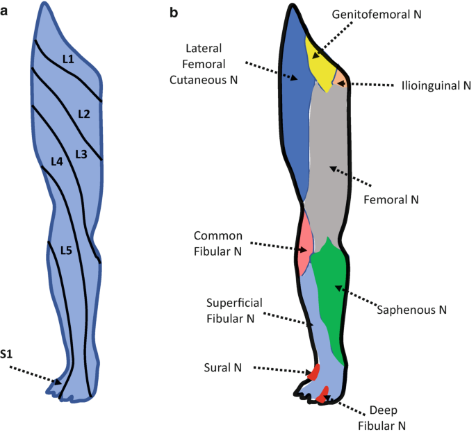
Source : link.springer.com
Dermatome Legs Royalty Free Images, Stock Photos & Pictures

Source : www.shutterstock.com
Dermatome Map of the Lower Limb Labeled Diagram Stock Illustration
Source : stock.adobe.com
Lower Limb Dermatomes Anatomy, How To Check Dermatomes?
Source : samarpanphysioclinic.com
Dermatome Map Lower Leg Dermatomes and Myotomes | Sensation | Anatomy Geeky Medics: The main skeletal muscles of the upper leg are: While the upper leg is the powerhouse, the lower leg is responsible for stability of the ankle and foot, Frankinburger says. The main skeletal muscles . This paper discusses the common causes of exercise induced lower leg pain, including chronic compartment syndrome (CCS), medial tibial stress syndrome (MTSS) and chronic calf tears (CCT). The .